Unfortunately, low back pain is one of the most common complaints in the United States with neck pain following closely behind it. With roughly 80% of individuals experiencing some form of low back pain in their lifetime, most of us have firsthand experience in back or neck pain. Although some patients only experience localized pain in their necks and low backs, it is very common for patients to complain of radicular symptoms or symptoms that travel into their upper or lower extremities. These symptoms can include, but are not limited to pain, numbness, tingling, burning, and muscle weakness. When symptoms are felt radiating into their extremities most patients say they have a “pinched nerve”. Although most individuals have heard about “pinched nerves,” they don’t always understand exactly what it is. Before we go further into discussing how a pinched nerve occurs, let’s take a brief look at the anatomy of our nerves.
The cervical spine consists of 7 vertebrae (C1-C7) which are between the skull and the thoracic spine. The lumbar spine consists of 5 vertebrae (L1-L5) which are between the thoracic spine and the sacrum (tail bone). In between every vertebra in the spine, there are intervertebral discs which act as shock absorbers and cushion the joints, allowing our weight to be distributed through the spine and provide support. The intervertebral disc is made up of 85-90% water, but as we age this amount decreases to 65% (2). Our nerves all start in our spinal cord. At every level of our spine, different nerves branch out of the cord on both the right and left side of the spinal column. A nerve is comprised of a group of axons that are enclosed by a connective tissue sheath. Each nerve throughout the cervical spine branches out and innervates the different muscles in our upper extremities. While the nerves exiting the lumbar spine branch out and innervate the muscles and tissues in our lower extremities.
How Does a Nerve Become Pinched?
When there is normal healthy movement of tissues throughout the body, the tissues will move in conjunction with our joints, allowing us to move pain-free. Nerve compression is often a result of a biomechanical issue resulting in static pressure on the nerve. This nerve compression (pinching) can occur anywhere along the length of the nerve.
It is common for the nerve to become compressed at the spinal level as a result of:
• Disc Herniation
• Spinal Degeneration/Spinal Stenosis – narrowing of the spinal canal
Or it can occur anywhere along the nerves pathway (peripheral nerve entrapment)
• tunnels where nerves have to pass (ex: carpal tunnel syndrome)
• areas where nerves branch/divide off
• areas where nerves are tethered down in the sheath

Two factors that affect the severity of nerve compression are the amount of load being applied to the nerve and the length of time the nerve is compressed. As the load and length of time increases, the nerve becomes aggravated and inflamed. The increase in inflammation results in inadequate mobility of the nerve, which can be quite painful. The greater the compression and inflammation around the nerve, the more the symptoms will radiate (spread). In the cervical spine, symptoms can be felt anywhere along the neck, shoulder, shoulder blade, forearm, and hand. In the lumbar spine, symptoms can occur in the back, buttocks, hip/groin, thigh, leg, and foot.
Tension is another contributing factor that will affect nerves. Unlike muscles that stretch, nerves tend to slide and glide within the connective sheath container that they lie within. Muscles are able to withstand increased tensile forces due to their higher extensibility. However, nerves do not stretch and do not respond well when there are increased tension forces applied. As tension is applied to a nerve, it is essentially being tugged on, which will cut off or reduce the blood supply to the nerve. Just like all tissues in our body, adequate blood supply is essential for oxygen delivery and ensures tissue viability.
Treatment Options
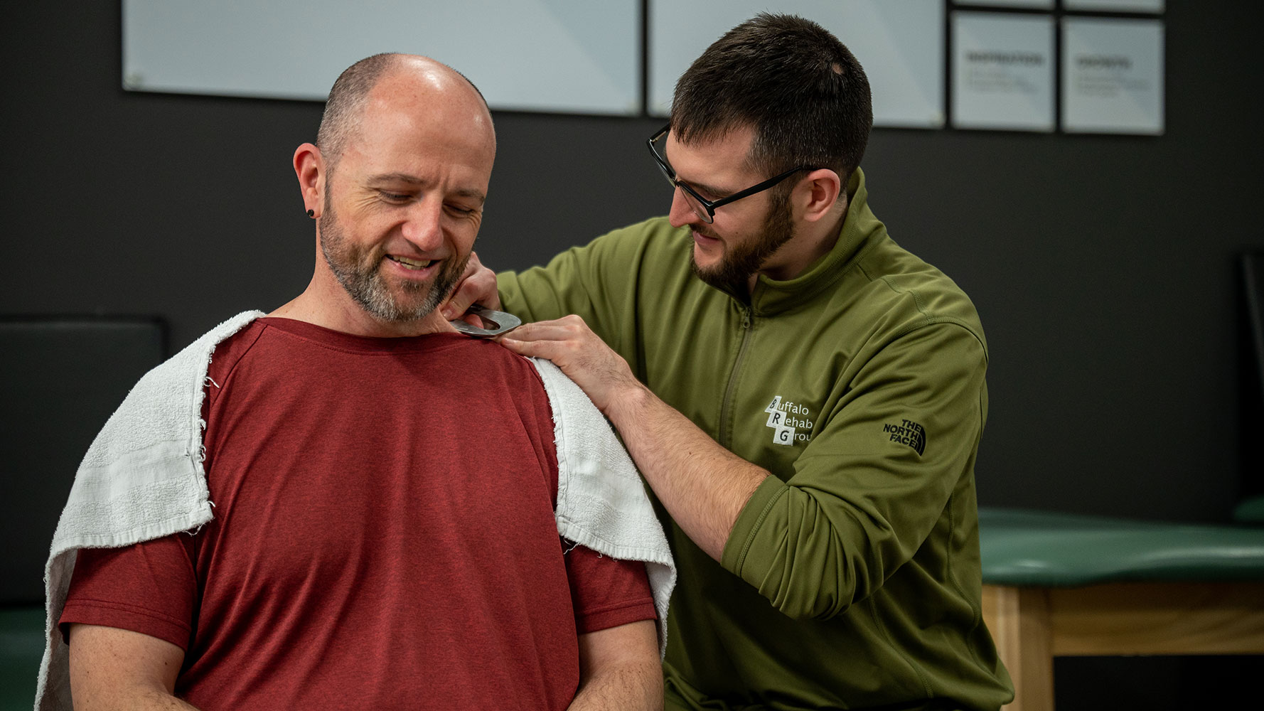
The location of symptoms and the information from a patient’s history will help the therapist or clinician determine which nerve and where the compression may be. Once a diagnosis is established, it is essential to provide the nerve and surrounding tissue an environment that will promote healing. We can do this by identifying specific lifestyle choices that may be contributing aggravating factors to reduce the load on your injured tissue. Gentle mobilizations and oscillatory movements can be performed manually to help free up and return mobility to the nervous system. A specific plan of care and home exercise program will be established by your Physical Therapist that will be tailored towards your symptoms and specific needs.
If you have symptoms that resemble nerve pain, click the link below to schedule your free discovery visit with a physical therapist at Buffalo Rehab Group.
References
1. Butler, David S. The Sensitive Nervous System. Noigroup Publications, 2008.
2. Magee, David J. Orthopedic Physical Assessment. Elsevier/Saunders, 2014.
3. Ellis, Richard F., and Wayne A. Hing. “Neural Mobilization: A Systematic Review of Randomized Controlled Trials with an Analysis of Therapeutic Efficacy.” Journal of Manual and Manipulative Therapy, vol. 16, no. 1, 2008, pp. 8-22.